Classification of Defects
Adapted from Mohan, R. et al. Aesthetic and functional facial transplantation: a classification system and treatment algorithm. Plast Reconstr Surg 133, 386-397 (2014).
Classifying the facial deficit helps physicians determine a treatment algorithm but also helps in communicating to other practitioners the nature of the deficit in standardized terms.
Soft-Tissue Defect
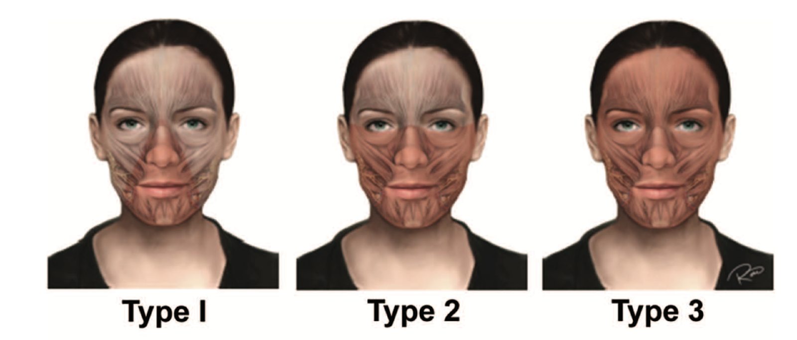
Type 1, the oronasal soft-tissue defect, includes loss of the upper or lower lips, commissure, and the nasal subunit structures (soft tissue, lining, or support). It involves the following functional impairments: oral sphincter, lip depression, and lip elevation. For a defect to be classified as type 1, it may contain a deficit of all or some of the structures listed. In the rare instance that there is an isolated defect of the oral subunit without involvement of the nasal subunit, this would be classified as type 0.
Type 2, the oronasal-orbital soft-tissue defect, includes all of the soft-tissue components of type 1 along with the inferior eyelids and cheek soft-tissue subunits. For a defect to be classified as type 2, it must comprise a deficit involving some or all of the soft tissue associated with the inferior orbital and cheek regions. Isolated defects of these regions would also be classified as type 2.
Type 3, the full facial soft-tissue defect, includes the soft-tissue defect of type 2 along with the upper eyelids and forehead. The superior border of this facial subunit is the anterior hairline and the lateral border is the preauricular region anterior to the tragus. For a defect to be classified as type 3, it must contain a deficit involving some or all of the soft tissues of the upper eyelid or frontal regions.
For all of the soft-tissue types described, the patient must have a defect comprising a significant portion of the subunit (>40 percent) in order to be classified as a particular type.
Hard-Tissue Defect

Type A includes defects of the maxillary Le Fort I segment as shown. For a bony defect to be classified as type A, it must contain a partial or complete defect of the maxilla but would be defined at a level cephalad to the dentition (i.e., the Le Fort I maxillary segment).
Type B involves the nasal bones, portions of the maxilla and zygoma, and inferomedial orbital bones associated with a Le Fort III osseous segment. Type B defects may also include the vomer, ethmoid, and medial orbits. The defect must contain the zygomatic-maxillary, nasal, and inferomedial orbital bones as well as the maxillary alveolus to be considered type B.
Type C includes the supraorbital bones and frontal bone associated with the segments of a monobloc osteotomy. Type C may involve the bones mentioned in the previous types, but it must contain a deficit in the bones superior to the ones in a Le Fort III segment and the maxillary alveolus.